Fig. 2.
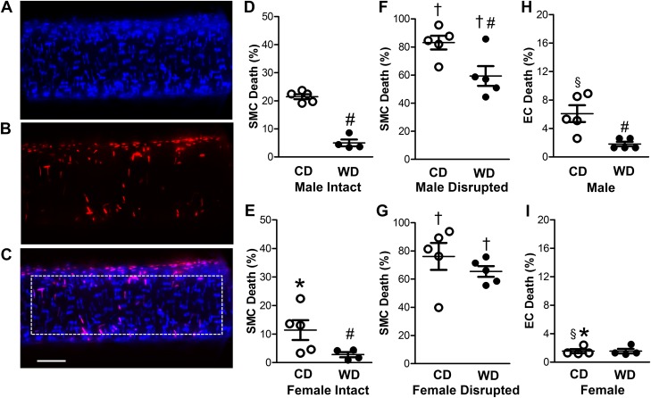
Female sex and Western-style diet (WD) protect against H2O2-induced cell death in the arterial wall. A: Hoechst 33342 staining of nuclei from all cells in the wall of a SEA. B: propidium iodide staining of nuclei of dead cells in vessel wall. C: merged image of A and B. Dotted rectangle indicates region of interest for cell counts, which shows 14 dead and 94 total SMC nuclei with 3 dead and 59 total EC nuclei. Scale bar = 50 µm (applies to all images). D–G: percentage of dead SMCs for SEAs from male intact (D), female intact (E), male endothelium-disrupted (F), and female endothelium-disrupted (G) SEAs of mice fed the control diet (CD) or WD following exposure to H2O2 (200 µM, 50 min). For intact vessels, SMC death was greater in males than females and was reduced in WD vs. CD for both sexes. Endothelial disruption increased SMC death in all cases, with the protective effect of the WD persisting in males. H and I: EC death of intact SEAs was lower in females (I) than males (H), and the protective effect of the WD was manifest in males but not females. Note difference in y-axis scales across D–H and E–I. Values are means ± SE; n = 4–5 vessels per group. *P < 0.05, female vs. male. #P < 0.05, WD vs. CD. †P < 0.05, endothelium-disrupted vs. intact. §P < 0.05, EC vs. SMC of endothelium-disrupted within sex.

