Fig. 4.

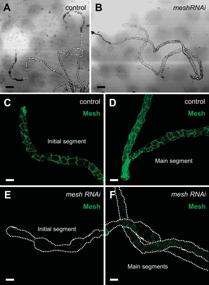
Developmental principal cell mesh knockdown in the larval Drosophila Malpighian tubule. A and B: bright-field images of the anterior Malpighian tubules from a nonwandering third-instar control larva (w;c42-GAL4/+; A) and developmental principal cell mesh knockdown larva (w;UAS-meshRNAi/+;c42-GAL4/+; B). The mesh knockdown tubules reveal some dilation compared with control tubules. C and D: Mesh immunolocalizes to the smooth septate junctions between the epithelial cells in control tubules. E and F: decreased Mesh immunofluorescence is observed in mesh knockdown tubules. Five samples per genotype were examined. Scale bars, 100 µm (A and B) and 50 μm (C–F).
