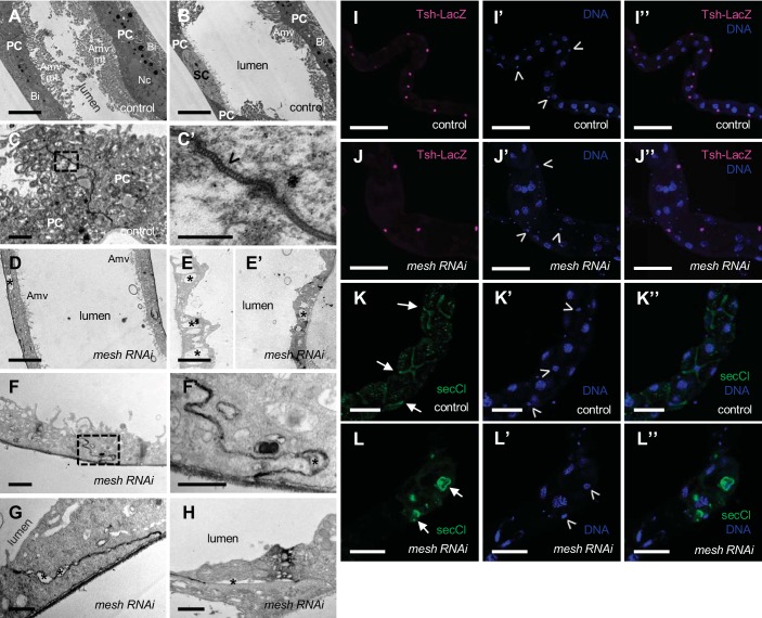
Fig. 7.
Developmental principal cell mesh knockdown in the Drosophila Malpighian tubule results in disruption of epithelial architecture and smooth septate junction (sSJ) organization. A–H: transmission electron micrographs of 1-day-old adult female fly anterior tubule main segment epithelial cells. Compared with a control tubule main segment (w;c42-GAL4/+; A and B), the epithelial cells of a principal cell mesh knockdown tubule main segment (w;UAS-meshRNAi/+;c42-GAL4/+; D–H) reveal a cytoplasm containing empty vacuoles (*), reduced or absent apical membrane microvilli (Amv) and associated mitochondria (mt), and reduced basal membrane infoldings (Bi). The sSJs between the principal cells (PCs) in a control tubule main segment (C and C′) show parallel plasma membranes and ladderlike septa (open arrowhead). C′ shows a higher-magnification image of dashed-box area in C. In mesh knockdown tubule main segment sSJs (F–H), the plasma membranes of adjacent cells are less parallel, and frequent large intercellular gaps are observed (*). F′ shows a higher-magnification image of dashed-box area in F. I–I′′ and J–J′′: confocal images of a 1-day-old adult female fly anterior tubule main segment expressing teashirt (tsh)-lacZ without (control; w;tsh04319/+;c42-GAL4/+) or with upstream activation sequence (UAS)-mesh RNA interference (mesh RNAi; w;tsh04319/+;c42-GAL4/UAS-meshRNAi) and stained for β-galactosidase (magenta) and DNA (blue, TOPO-3). Both control and mesh knockdown tubules express Tsh-LacZ restricted to the stellate cells with smaller nuclei (open arrowheads in I′ and J′). K–K′′ and L–L′′: confocal images of a 1-day-old adult female fly anterior tubule main segment stained for secretory chloride channel (secCl, green) and DNA (blue, DAPI). Control (w;c42-GAL4/+; K–K′′) and mesh knockdown (w;UAS-meshRNAi/+;c42-GAL4/+; L–L′′) tubules show secCl expression in the stellate cells with smaller nuclei (arrows and open arrowheads in K, K′, L, and L′). However, compared with control tubule main segment, the stellate cells of mesh knockdown tubules are abnormally shaped. Nc, nucleus; SC, stellate cell. Three samples per genotype were examined in A–J; 10 samples per genotype were examined in K and L. Scale bars, 100 µm (I–I′′ and J–J′′), 50 µm (K–K′′ and L–L′′), 5 μm (A, B, D, E, and E′), 1 μm (C, F, G, and H), 500 nm (F′), and 200 nm (C′).

