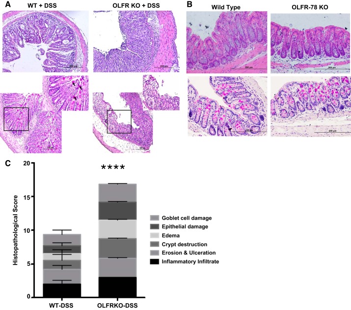
Fig. 4.
Structural changes in colons of wild-type (WT) and olfactory receptor-78 (Olfr-78) KO mice treated with dextran sodium sulfate (DSS). A: formalin-fixed distal colon tissues from WT and Olfr-78 knockout (KO) mice treated with DSS were subjected to H&E staining (top) assessing structural changes and Periodic acid-Schiff staining (PAS; bottom) for mucin production by goblet cells. Boxed area in black borders is zoomed in on the top right corner to show mucin staining (n = 3 for WT and 6 for KO). B: same as in A except formalin-fixed distal colon tissues from WT and Olfr-78 KO mice not treated with DSS were used. C: histological scores from H&E- and PAS-stained sections are presented. ****P < 0.0001.

