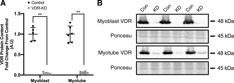
Fig. 1.
Generation of vitamin D receptor (VDR) loss of function C2C12 myoblasts. A: quantification of VDR protein content in VDR-knockdown (KD) compared with control myoblasts and myotubes. B: representative immunoblot images of VDR protein content in VDR-KD myoblasts and myotubes. **P < 0.005, independent t test. Data are means ± SD (n = 5-6 lanes/group) and represented as a fold change from control.

