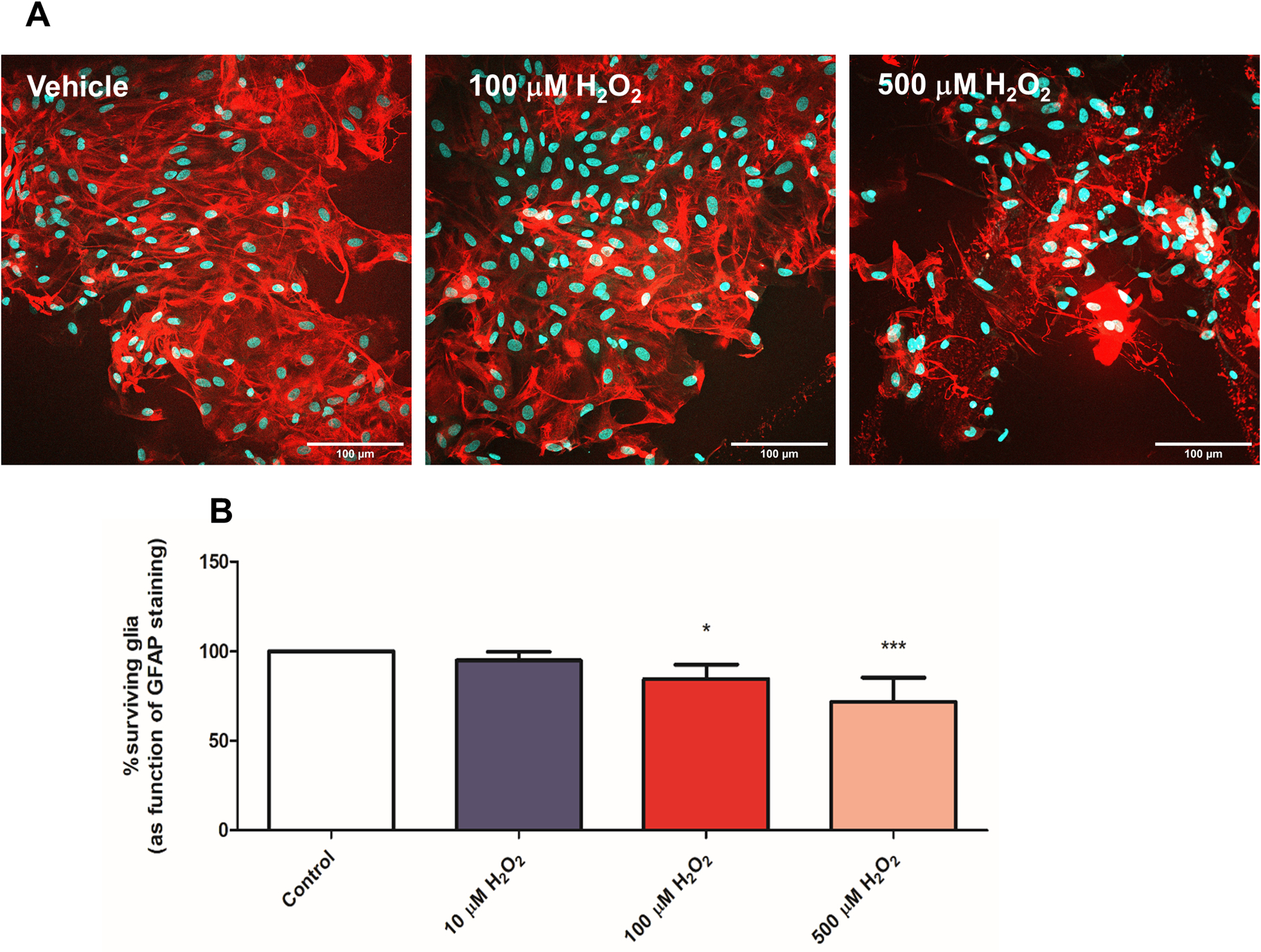
Figure 5. H2O2 -mediated oxidative stress results in significant glial cell death.

A. Representative images of pure glia cultures incubated with vehicle (control), 100 and 500μM H2O2 for 20 min, indicating loss of glia due to the insults.
B. Quantification of GFAP+ cells indicates 100 μM and 500μM H2O2 insults result in significant glial cell death in pure glial cultures. Survival quantification was determined by comparing GFAP staining to total DAPI staining and cultures were normalized to control, vehicle-treated cultures. 3 coverslips were assessed per treatment condition per experiment. Neuronal survival data is represented as the mean ± SEM of 2 independent experiments (n=6) and control levels were not statistically different for normalization purposes. ***p<0.001, *p<0.05, vs. control (no insult).
