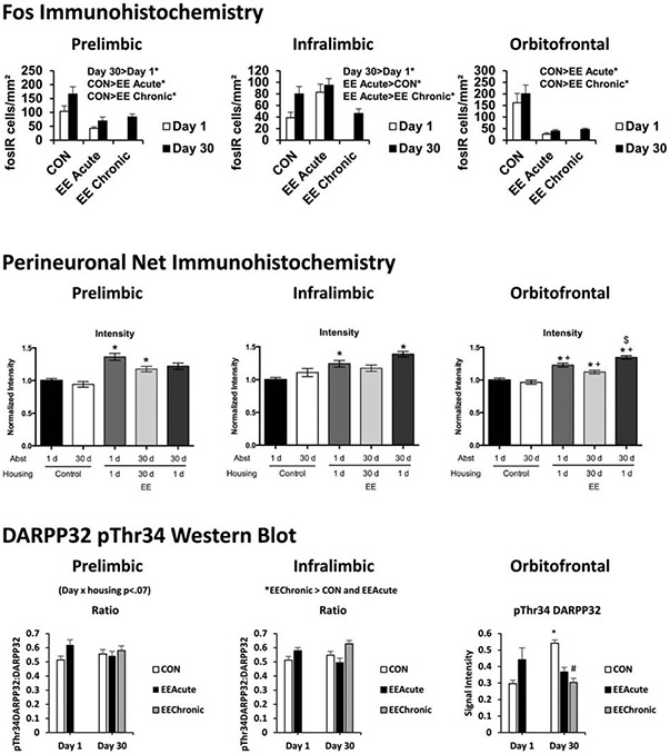
Fig. 9. EE alters synaptic plasticity and dopamine-related signaling in meso-cortico-limbic terminals in rats with a history of sucrose self-administration.
Selected molecular mapping results: A comparison of effects in the prelimbic, infralimbic, and orbitofrontal cortices. Top: cFos expression assessed using immunohistochemistry. Middle: PNN intensity assessed using immunohistochemistry. Bottom: DARPP32 phosphorylation at Thr34 assessed using Western blot. Top: * indicates significant difference (comparisons depicted above each panel), Middle: * indicates significant difference from Day 1 Controls; + indicates significant difference from Day 30 Controls; $ indicates significant difference from Day 30 Chronic EE, Bottom: * indicates significant difference from Day 1 for that housing condition or as otherwise depicted above each panel; # indicates significant difference from CON on that day of abstinence; P < .05. Means ± SEMs indicated on figures. Top figure reproduced from “Effects of acute or chronic environmental enrichment on regional Fos protein expression following sucrose cue-reactivity testing in rats,” by J.W. Grimm, J.L. Barnes, J. Koerber, E. Glueck, D. Ginder, J. Hyde, and L. Eaton, 2016, Brain Structure and Function, 221, p. 2825. Copyright 2015 by Springer-Verlag Berlin Heidelberg. Figure adapted with permission. Middle figure reproduced from “Impact of environmental enrichment on perineuronal nets in the prefrontal cortex following early and late abstinence from sucrose self-administration in rats” by M. Slaker, J. Barnes, B.A. Sorg, and J.W. Grimm, 2016, PLoS One, 11:e0168256, Copyright 2016 by the authors. Bottom figure reproduced from “Sucrose abstinence and environmental enrichment effects on mesocorticolimbic DARPP32 in rats” by J.W. Grimm, E. Glueck, D. Ginder, J. Hyde, K. North, and K. Jiganti, 2018, Scientific Reports, 8:13174. Copyright 2018 by Springer Nature under the terms of the Creative Commons, creativecommons.org/licenses/by/4.0/

