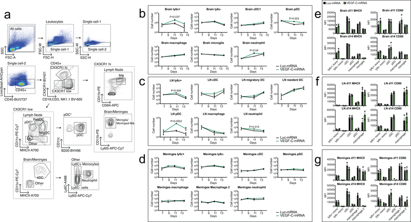
Extended Data Fig 8. Flow cytometry analysis of myeloid cell populations after VEGF-C treatment.
Mice bearing 7 day-tumors were treated with Luc-mRNA or VEGF-C-mRNA and evaluated for changes in myeloid populations. a Gating strategy for different myeloid cells. b-d Cell counts of different cell types were measured at different time points after VEGF-C-mRNA treatment. e-g MHCII and CD80 MFI levels were graphed and showed no significant alteration after VEGF-C-mRNA treatment. b and e are leukocytes from brain tissue. c and f are leukocytes from draining cervical lymph nodes. d and g are leukocytes from meninges (n = 3, 3 animals pooled for each replicate). Data are mean ± S.E.M *P < 0.05; **P < 0.01; ***P <0.001; ****P<0.0001 (two-tailed unpaired Student’s t-test)

