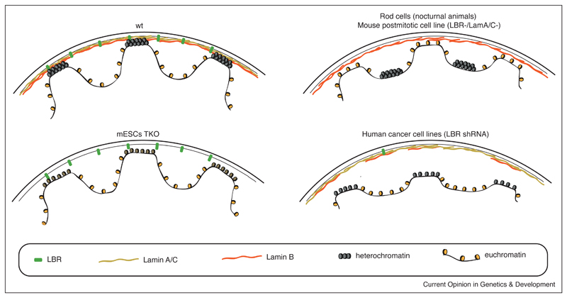
Figure 1. Schematic representation of lamina composition impact on chromatin conformation.
On the top left is a wild-type nucleus consisting of a nuclear lamina containing LBR and all lamins. The compacted heterochromatin is located at the nuclear periphery, while euchromatin is located more at the nuclear interior. In triple knockout mESCs that do not express any lamins (bottom left), genome–NL interactions are maintained but cLAD chromatin is decondensed [19••]. Rod cells of nocturnal animals that naturally lack LBR and Lamin A/C and mouse postmitotic cells in which LBR and Lamin A/C were mutated show inverted chromatin organization with heterochromatin localizing at the nuclear interior and euchromatin localizing at the periphery (top right) [23,25]. In human cancer lines in which LBR is downregulated, Lamin B1 expression is also affected and results in the detachment of heterochromatin from the NL (bottom right) [26••].

