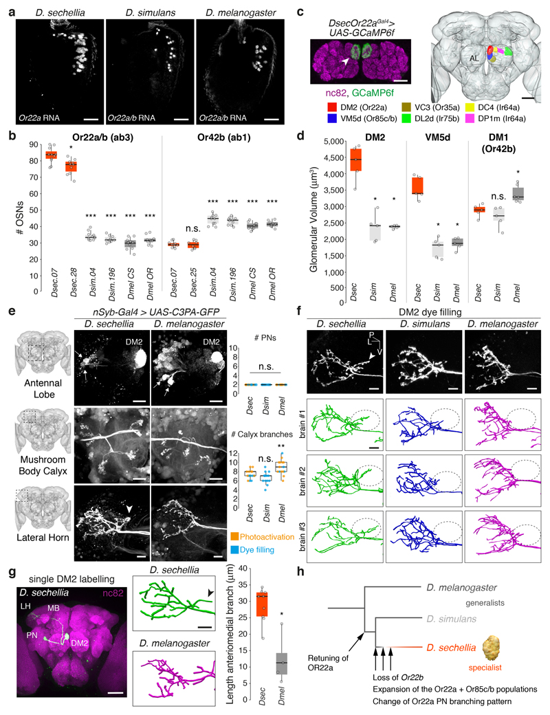
Fig. 4. Neuroanatomy of noni-sensing olfactory pathways.
a, Antennal Or22a/b RNA expression in different species. Scale bars, 25 μm.
b, Quantification of Or22a/(b) or Or42b OSNs (n = 8-11, females). Comparisons to Dsec.07 are shown (pairwise Wilcoxon rank-sum test, P values adjusted for multiple comparisons using the B&H method, b, d, e): *** P < 0.001; ** P < 0.01; * P < 0.05; n.s. P > 0.05.
c, Left: Or22aGal4-driven GCaMP6f expression in DM2 (arrowhead); neuropil visualised with nc82 (magenta). Scale bar, 25 μm. Right: Antennal lobe (AL) glomerular segmentation in D. sechellia (Extended Data Fig. 3). Scale bar, 50 μm.
d, Quantification of DM2, VM5d and DM1 volumes (n = 5 females).
e, DM2 projection neurons (PNs) labelled via photoactivation in D. sechellia (DsecnSyb-Gal4/UAS-C3PA-GFP) and D. melanogaster (UAS-SPA-GFP/UAS-C3PA-GFP;nSyb-Gal4/UAS-C3PA-GFP). Left: Image acquisition site. Top: AL with labelled PNs (arrows) and DM2 glomerulus; scale bar, 20 μm. Middle and bottom: PN innervation of mushroom body calyx and lateral horn (LH); scale bars, 10 μm. Arrowhead (e, f, g): extra anterio-medial branch in D. sechellia. Right: quantification of DM2 PNs (top) and calyx branches (bottom) (n = 14-17 females).
f, LH arbours of dye-filled DM2 PNs. Genotypes: D. sechellia, D. melanogaster as in e, D. simulans: DsimOr22aGFP. Below: representative LH DM2 arbour traces. Ovals: location of D. sechellia-specific branch. P, posterior; L, lateral; V, ventral. Scale bars, 10 μm.
g, Left: single dye-filled DM2 PN in D. sechellia; scale bar, 50 μm. Middle: representative LH arbour traces of DM2 PNs in D. sechellia and D. melanogaster; scale bar, 10 μm. Right: quantification of anteriomedial branch length (n = 4-9 females) (pairwise Wilcoxon rank-sum test): * P < 0.05.
h, Evolution of structural and physiological changes in the Or22a pathway.

