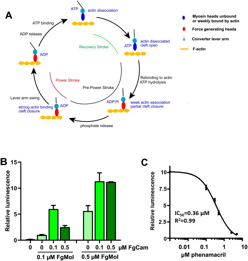
Fig 1. FgMyoI ATPase activity.
(A) Myosin/actin catalytic cycle (modified from [13]). ATP-bound myosin is dissociated from actin and becomes rapidly hydrolyzed in the catalytic site. Release of the hydrolyzed phosphate is slow and requires actin binding for acceleration. Initial weak electrostatic actin binding partially closes the actin-binding cleft and stabilizes a switch loop movement. This allows concerted phosphate release, full cleft closure, actin binding and lever arm movement (power stroke). This is followed by ADP release, which allows rapid ATP rebinding, actin dissociation, and a lever arm recovery stroke. (B) Calmodulin dependence of the FgMyoI activity measured by ATP-Glo ATPase luminescence assay. (C) Phenamacril inhibits the ATPase activity of FgMyoI; n = 3, error bars = SD.

