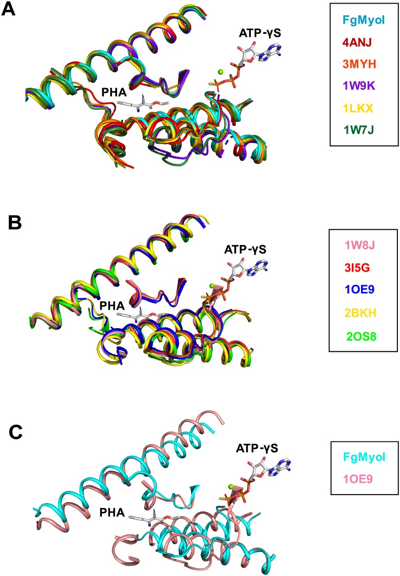
Fig 7. Alignment of myosin domains in nucleotide-bound and nucleotide-free states.
(A) Alignment of FgMyoI with five structures of myosin motor domains in nucleotide-bound state with relatively more open actin-binding clefts. (B) Alignment of five structures of myosin motor domains in nucleotide-free state with closed actin-binding clefts. The positions of phenamacril (PHA) and ATP-γS from FgMyoI are shown for orientation. Parts of the motor domains that are not close to the phenamacril-binding pocket have been removed for clarity. (C) Close-up of structure overlay of FgMyoI with that of chicken myosin in nucleotide-free, closed cleft conformation. (4ANJ [44]/2BKH [45]: Sus scrofa class 6 myosin; 3MYH [46]/1W9K: Dictyostelium discoideum class 2 myosin; 1LKX [36]: Dictyostelium discoideum class 1 myosin; 1W7J [40]/1W8J [40]/1OE9 [41]: Gallus gallus class 5 myosin; 3I5G [42]: Doryteuthis pealeii class 2 myosin; 2OS8 [42]: Placopecten magellanicus class 2 myosin).

