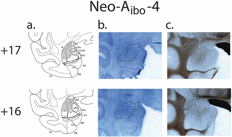
Figure 1.
Amygdala lesion extent for a single case (Neo-Aibo-4): Atlas drawing outlining the extent to damage in grey (a), Corresponding photomicrogrpahs of 50μm coronal sections of the left amygdala stained for cell bodies with Nissl (b), and fiber tracts with silver impregnation (c). Dashed lines on the Nissl photomicrographs outline the borders of the cell loss (b).

