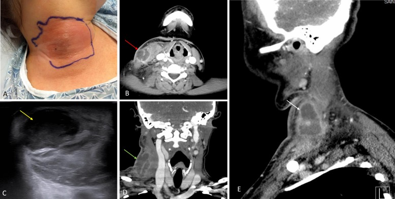
Figure 1.
(A) Clinical picture showing swelling of the neck at the site of injection. (B) CT neck axial view showing loculated pus collection. (C) Ultrasonography (USG) of the neck confirmed a hypoechoic spherical collection of echogenic fluid with well-defined borders. (D, E) CT coronal view (D) and sagittal view (E) confirming the abscess in the sternocleidomastoid muscle measuring 2.6×2.6×5.3 cm.

