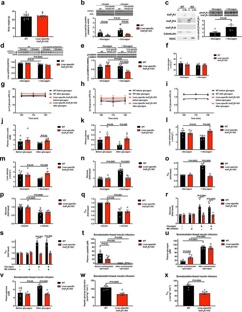
Extended Data Figure 1.
Glucagon acutely stimulates hepatic gluconeogenesis by increasing hepatic acetyl-CoA content and pyruvate carboxylase flux. (a) Body weight (n=11). (b) Liver InsP3R-I protein expression (n=5). Blots in Figures 1f and 2a, and Extended Data Figures 1b, 1d, 1f, 3f, 3g, and 4a were stripped and re-probed for all proteins of interest. *P<0.05 vs. InsP3R-I KO mice not treated with glucagon. (c) InsP3R protein expression in cytosolic (“c.”) and crude mitochondrial (“c.m.”) fractions from primary hepatocytes, in which VDAC was examined as a marker for mitochondrial protein content, and calreticulin as a marker for non-mitochondria-associated membrane protein. On the right, Liver InsP3R-I phosphorylation in mice infused with glucagon (n=5). The blot for total InsP3R-I is duplicated from Extended Data Figure 1b. (d) Liver CRTC2 phosphorylation (n=5). The CRTC2 phosphorylation gel was stripped and re-probed to assess HSP90 (loading control). (e) Liver CAMKIV phosphorylation ± a 2 hr acute infusion of glucagon (n=5). (f) Liver glycogen content (n=5 WT–glucagon, otherwise n=6). No differences were observed using one-way ANOVA with Bonferroni’s multiple comparisons test. (g)-(i) Plasma [m+1], [m+2], and [m+7] plasma glucose enrichment during a 120 min infusion of [3-13C] lactate and [2H7] glucose (n=5 WT and 6 KO, with the exception of panel (i), in which n=4 WT+glucagon at 100 and 110 min). (j)-(k) Plasma total amino acid and alanine concentrations (n=5 WT and 6 KO). In panels j-k, groups were compared before and after glucagon by the 2-tailed paired Student’s t-test, and genotypes were compared by the 2-tailed unpaired Student’s t-test. (l)-(m) Liver total amino acid and alanine concentrations (n=5). (n)-(o) In vitro glucose production (n=9) and VPC flux (n=4) in isolated hepatocytes. (p)-(q) In vitro glucose production (n=9) and VPC flux (n=4) in isolated hepatocytes with and without 150 pM insulin. Basal data (no insulin) are duplicated from panels n and o. (r)-(s) In vitro glucose production (n=8) and VPC flux (n=3) in isolated hepatocytes with and without a malic enzyme inhibitor. **P<0.01, ****P<0.0001 vs. WT-glucagon-ME inhibitor. (t)-(v) Plasma c-peptide, glucagon, and glucose concentrations in mice (n=6 WT and 7 KO) treated with somatostatin, basal insulin, and glucagon. Comparisons before and after glucagon used the 2-tailed paired Student’s t-test. (w)-(x) Endogenous glucose production and VPC (n=6 WT and 7 KO). In all panels, unless otherwise stated, comparisons with and without glucagon, insulin, or malic enzyme inhibitor, and WT vs. KO were performed using the 2-tailed unpaired Student’s t-test. In all panels where comparisons were performed (i.e. all panels with the exception of g-i), if no p-value is shown, groups were not significantly different. In all panels, the mean±S.E.M. are shown.

