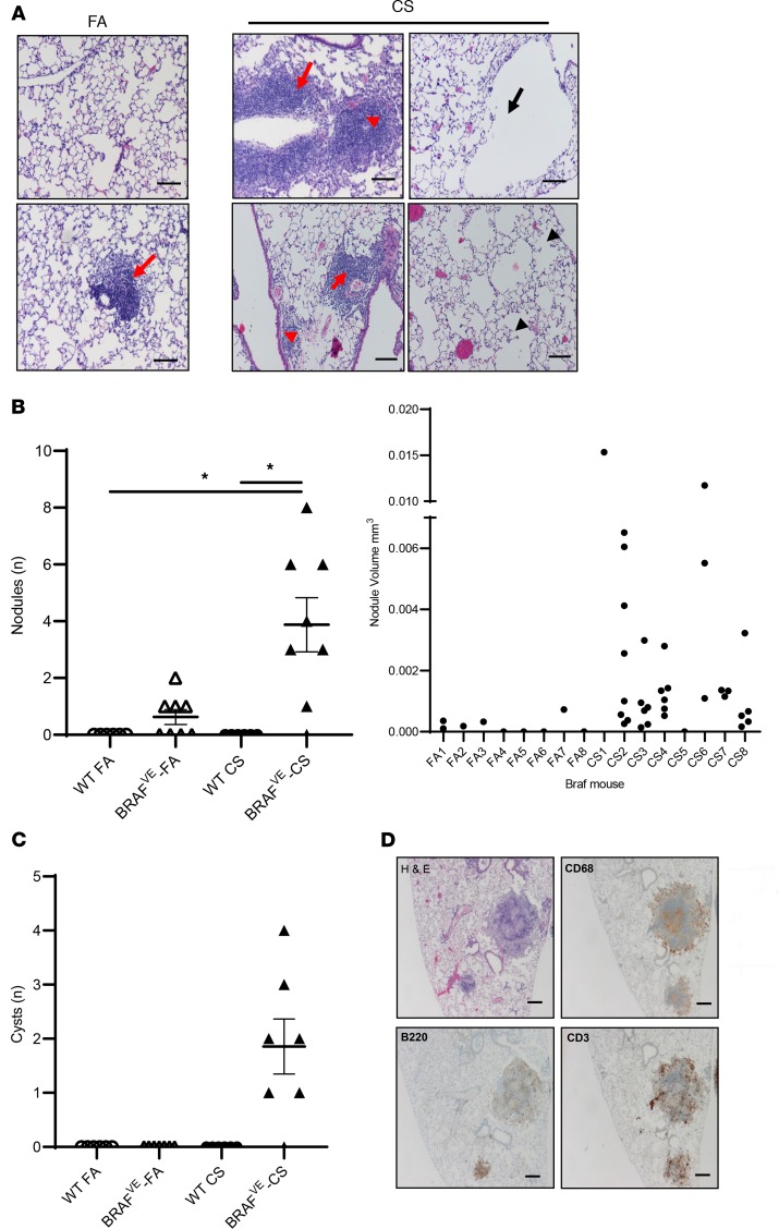
Figure 1. Exposure of mice with BRAF-V600E expression in CD11c+ DCs to CVS results in PLCH-like lesions.
(A) Representative H&E-stained sections showing lung pathology in BRAFVE mice exposed to FA or CS for 4 months. Red arrowheads indicate parenchymal nodule formation and red arrows indicate perivascular and peribronchiolar cell infiltration. Black arrows indicate cyst-like lesions and black arrowheads indicate areas of heterogeneous alveolar tissue destruction. Scale bar: 100 μm. Representative image of n = 8 mice per group. (B) Number and volume of pulmonary nodules. Nodules were identified as large inflammatory cell infiltrates not surrounding an airway or vessel. The volume of each individual nodule was estimated as described in Methods. n = 8 mice per group. (C) Enumeration of pulmonary cystic structures more than 200 μm in diameter. Representative lesions from n = 8 mice per group. (D) Representative IHC staining of nodular inflammatory lesions in lungs of BRAFVE mice exposed to CS for 4 months. Scale bar: 250 μm. Representative lesions from n = 8 mice per group. For experiments shown, 1-way ANOVA with Tukey’s multiple-comparisons analysis was performed. *P < 0.05.

