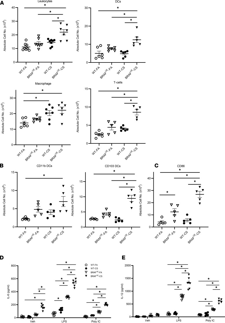
Figure 2. Increased inflammatory cells and disrupted DC homeostasis in the lungs of BRAFVE mice.
(A) Absolute cell numbers of leukocytes, DCs, macrophages, and T cells in the dissociated lungs of WT (n = 5–8 mice per group) and BRAFVE (n = 5–7 mice per group) mice were determined by flow cytometry. Leukocytes were identified as CD45+; DCs were identified as CD11c+, MHC II+, and autofluorescencemid/low cells; macrophages were identified as CD11c+ and autofluorescencehi cells; and T cells were identified as CD11c– and CD3+cells (n = 5 mice per group). (B) The absolute numbers of CD11b+ DCs and CD103+ DCs were determined by flow cytometry. Both subsets were gated from the DC population. (C) The number of DCs expressing maturation marker CD86 was determined by flow cytometry (n = 5 mice per group). (D and E) DCs were isolated from lungs and treated with or without 20 ng/mL IFN-γ for 2 hours before being treated with 1 μg/mL poly(I:C) or 1 μg/mL LPS for 16 hours. The supernatant was collected and IL-6 and IL-12 p40 were measured by ELISA (n = 5–7 mice per group). For experiments shown, ANOVA (1 way in A and B, 2 way in D and E) with Tukey’s multiple-comparisons analysis was performed. *P < 0.05. Data represent mean ± SEM.

