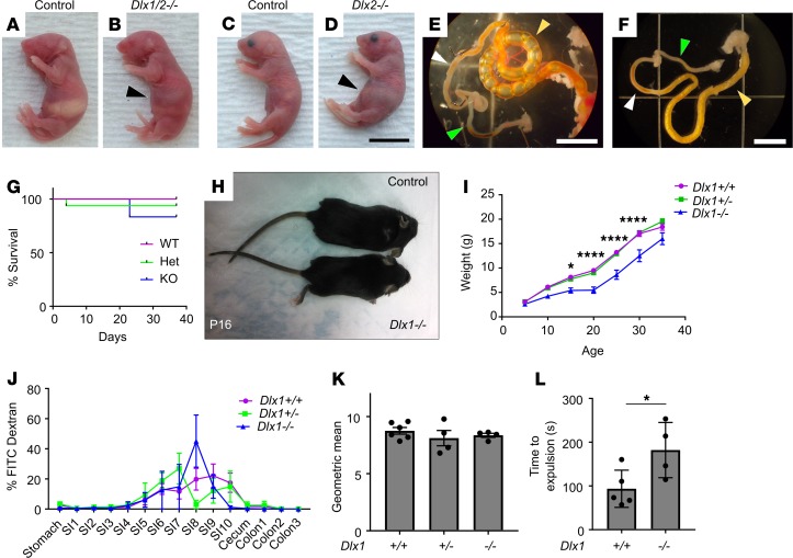
Figure 1. Dlx1/2–/– and Dlx2–/– mice have obvious abdominal distention at P0 due to bowel air accumulation, while Dlx1–/– mice exhibit poor weight gain but normal small intestine (SI) transit.
(A–D) Dlx1/2–/– (B) and Dlx2–/– (D) mice exhibited dramatic bowel distention (black arrowhead) compared to WT littermates (A and C). Scale bar: 1 cm. (E) Bowel of Dlx2–/– mouse viewed under dissecting microscope. Note air-filled proximal SI (yellow arrowhead) with absence of air in distal SI (white arrowhead) and colon (green arrowhead). Scale bar: 5 mm. (F) Control bowel for comparison. Scale bar: 5 mm. (G) Kaplan-Meier curve indicates most Dlx1–/– mice survived past 1 month of age (P = 0.4665; log-rank Mantel-Cox; n = 9 [+/+], 16 [+/–], 6 [–/–]). (H and I) Dlx1–/– mice were small and weighed significantly less than their WT littermates at P15 (P = 0.0433), P20 (P < 0.0001), P25 (P < 0.0001), and P30 (P < 0.0001); n = 9 [+/+], 14 [+/–], and 5 [–/–]; 2-way repeated-measures ANOVA. We include only mice that lived to P35. (J) Percentage FITC-dextran in distinct bowel regions 1 hour after oral FITC-dextran administration. SI1 to SI10 indicate sequential SI segments. (K) Weighted average of FITC bowel transit showed normal SI transit times in Dlx1–/– mice (P = 0.5182; 1-way ANOVA, n = 6 [+/+], 4 [+/–], and 4 [–/–]). (L) Dlx1–/– mice had slight delays in expulsion of a glass bead from distal colon compared with WT controls (P = 0.0399, Student’s t test, n = 5 [+/+] and n = 4 [–/–]). *P < 0.05; ****P < 0.0001.

