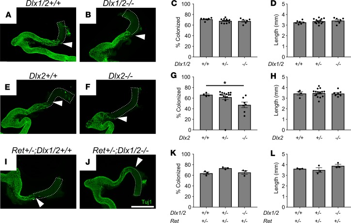
Figure 3. Dlx1/2–/–, Dlx2–/–, and Ret+/– Dlx1/2–/– mice have normal bowel colonization rates by ENCDCs or minimal defects at E12.5. (A and B) Confocal Z-stacks of WT (A) and Dlx1/2–/– (B) E12.5 bowel stained with TuJ1 antibody that labels early and mature neurons (green).
Maximum intensity projections are shown. White triangles mark distal-most TuJ1+ neuron processes. Scale bar: 1 mm. (C and D) Percentage hindgut colonized relative to total colon length (C; 1-way ANOVA, n = 7 [+/+], n = 13 [+/–], n = 6 [–/–]) and colon length (D; 1-way ANOVA, n = 7 [+/+], n = 13 [+/–], n = 6 [–/–]) were normal in Dlx1/2–/– mutants. (E–H) Representative WT (E) and Dlx2–/– (F) E12.5 bowel labeled with TuJ1 antibody. Dlx2–/– mice had a slight delay (66.5 ± 1.5% colonized [+/+] vs. 47.4 ± 5.2% colonized [–/–]) in ENS migration of unknown functional significance (G; P =0.0499; Kruskal-Wallis test with Dunn’s multiple-comparisons test, n = 4 [+/+], n = 15 [+/–], n = 7 [–/–]) and normal colon length (H). (I–L) Representative Ret+/– Dlx1/2+/+ (I) and Ret+/– Dlx1/2–/– (J) E12.5 bowel labeled with TuJ1 antibody. Dlx1/2–/– mice on a Ret-heterozygous background had normal extent of bowel colonization (I; 1-way ANOVA, n = 3 per genotype) and normal colon length (L; 1-way ANOVA, n = 3 per genotype). Scale bar: 1 cm. *P < 0.05.

