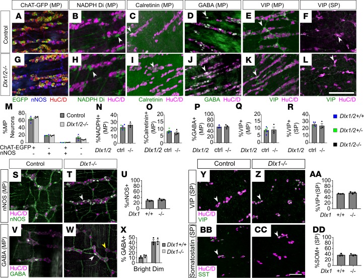
Figure 6. Dlx1/2–/– P0 and Dlx1–/– adult mice have normal ratios of neuronal subtypes.
(A–L) Representative images of HuC/D (red), nNOS (blue), and ChAT-EGFP (green; A and G), HuC/D (magenta) and NADPH-diaphorase (green; B and H), HuC/D (magenta) and calretinin (green; C and I), HuC/D (magenta) and GABA (green; D and J), HuC/D (magenta) and VIP (green; E, F, K, and L) immunostaining in P0 control and P0 Dlx1/2–/– bowel. SP, submucosal plexus; MP, myenteric plexus. (M–R) We observed no difference in ratios of myenteric ChAT-EGFP or nNOS+ (M), myenteric NADPH+ (N), calretinin+ (O), GABA+ (P), VIP+ (Q), or submucosal VIP+ (R) neurons to total HuC/D+ neurons in Dlx1/2–/– mice compared to control mice. WT and Dlx1/2+/– were grouped as “control” and color coded Dlx1/2+/+ (blue), Dlx1/2+/– (green) (Student’s t test, n = 3–6 per condition). (S, T, V, W, Y, Z, BB, and CC) Representative images of HuC/D (magenta) and nNOS (green; S and T), HuC/D (magenta) and GABA (green; V and W), HuC/D (magenta) and VIP (green; Y and Z), and HuC/D (magenta) and somatostatin (green; BB and CC) immunostaining in adult control and Dlx1–/– ENS. SP, submucosal plexus; MP, myenteric plexus. (U, X, AA, and DD) We observed no difference in myenteric nNOS+ (U) or GABA+ (X) neurons, and no difference in submucosal VIP+ (AA) or somatostatin+ (DD) neurons in Dlx1–/– ENS (Student’s t test, n = 3 per condition). Arrowheads indicate examples of neurons that were counted. For GABAergic neurons in Dlx1–/– adults, white arrowheads indicate brightly positive GABAergic neurons, while yellow arrowheads indicate dimly positive GABAergic neurons. Scale bars: 100 μm.

