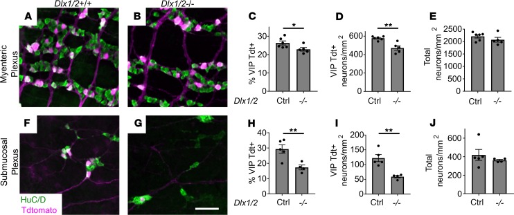
Figure 8. Vip-lineage TdTomato+ neuronal numbers are decreased in Dlx1/2–/– myenteric and submucosal plexus.
(A and B) Myenteric plexus from Dlx1/2+/(+or–) VIP-IRES-CreCre/wt R26R-TdTomato+ control (A) and Dlx1/2–/– VIP-IRES-CreCre/wt R26R-TdTomato+ mouse (B) stained with ANNA-1 antibody labeling HuC/D (green) reveals fewer TdTomato+ cells in mice lacking Dlx1/2. (C and D) Quantification of A and B shows a reduced proportion of TdTomato+ neurons in Dlx1/2–/– myenteric plexus (C; P = 0.047, n = 6 [ctrl], n = 5 [–/–]) and reduced overall TdTomato+ neuronal density (D; P = 0.0017, n = 6 [ctrl], n = 5 [–/–]). (E) Total MP neuronal density was unchanged in Dlx1/2 mutant mice (E; P = 0.26, n = 6 [ctrl], n = 5 [–/–]). (F and G) Dlx1/2–/– VIP-IRES-CreCre/wt R26R-TdTomato+ mutants also have fewer TdTomato+ (red) HuC/D+ cells in their submucosal plexus. (H and I) Quantification of F and G showing decreased proportion of TdTomato+ neurons relative to total (H; P = 0.0098, n = 5 [ctrl], n = 4 [–/–]) and decreased TdTomato+ neuronal density (I; P = 0.0027, n = 5 [ctrl], n = 4 [–/–]). (J) Total submucosal plexus neuronal density was unchanged in Dlx1/2–/– mice. Scale bar: 100 μm. *P < 0.05, **P < 0.001.

