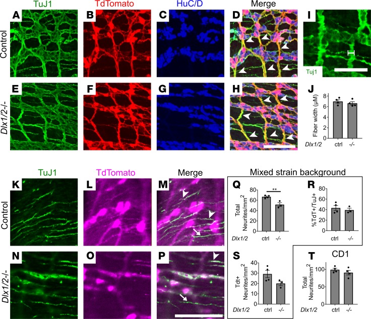
Figure 9. Dlx1/2–/– mice have apparently normal large nerve fiber bundles but decreased CSM neurite density on some strain backgrounds.
(A–H) Myenteric plexus labeled with TuJ1 (green; A and E), TdTomato (red; B and F), and HuC/D (blue; C and G). All large nerve fiber bundles (white arrowheads) were TdTomato+. (I and J) Myenteric plexus nerve fiber bundle width (white line, I) was similar in Dlx1/2–/– and control mice (J; P = 0.4565, Student’s t test, n = 4 per genotype). (K–P) Circular muscle neurites labeled with TuJ1 (green; K and N) and TdTomato (magenta; L and O) from P0 bowel. Some neurites were TdTomato+ and TuJ1+ (white arrowheads), while others were TuJ1+ alone (white arrows). (Q) Density of total TuJ1+ neurites was decreased in Dlx1/2–/– Vip-IRES-Cre TdTomato P0 bowel (P = 0.0077, Student’s t test, n = 4 [ctrl], n = 3 [–/–]). (R) Ratio of TdTomato+ neurites to TuJ1+ neurons was unchanged (P = 0.5587, Student’s t test, n = 4 [ctrl], n = 3 [–/–]). (S) Density of TdTomato+ neurites was not statistically different for control and Dlx1/2–/– mice (P = 0.1006, Student’s t test, n = 4 [ctrl], n = 3 [–/–]). (T) Density of total TuJ1+ neurites was normal for Dlx1/2–/– mice on a CD1 background (P = 0.3193, Student’s t test, n = 4 [ctrl], n = 4 [–/–]). Scale bars: 100 μm (D–H and K–P) and 50 μm (I). **P < 0.001.

