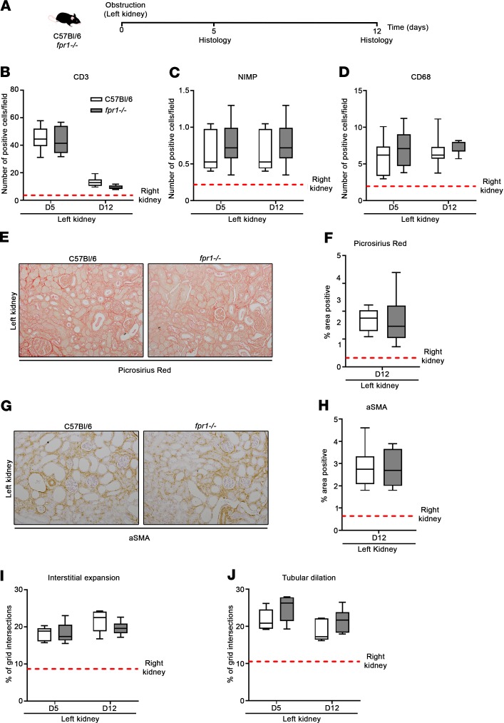
Figure 6. fpr1–/– mice are not protected from fibrosis in the unilateral ureteral obstruction model of kidney injury.
(A) The left kidney of C57BL/6 and fpr1–/– mice was obstructed by ligation of the corresponding ureter. The right kidney underwent the same surgical procedure without ligation. Mice were killed on days 5 and 12 after surgery and kidney tissue harvested. Number of (B) CD3+, (C) NIMP+, and (D) CD68+ cells per field (original magnification, ×20). Data represent the mean value of n = 20 randomly selected, nonoverlapping fields per mouse. Representative (E) Picrosirius red–stained and (G) α-smooth muscle actin–stained (αSMA-stained) kidney tissue. Percentage area positive of (F) Picrosirius red and (H) αSMA staining. Data represent the mean value of n = 20 randomly selected, nonoverlapping fields (original magnification, ×10). (I) Interstitial expansion and (J) tubular dilation as a percentage of grid intersections (excluding glomeruli) overlaying interstitial areas and tubular lumine. Data represent the mean value of n = 20 randomly selected, nonoverlapping fields (original magnification, ×20). n = 6–7 mice per group. No significant difference was seen between the right kidneys of C57BL/6 and fpr1–/– mice, and therefore mice were pooled and presented as mean (red-hashed line). Data were analyzed using a Mann-Whitney U test and presented as box-and-whisker plots. P > 0.05.

