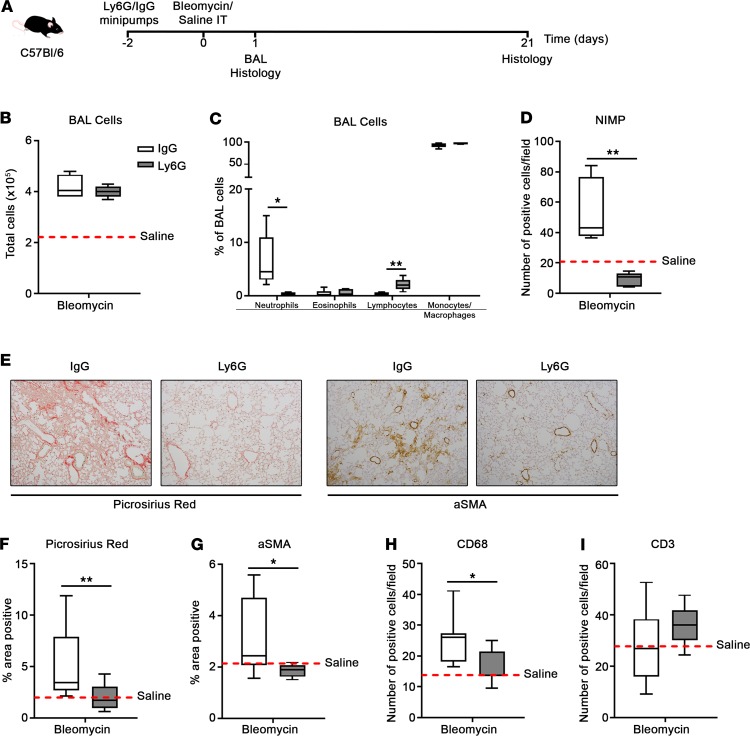
Figure 8. Neutrophil depletion attenuates bleomycin-induced lung fibrosis.
(A) Osmotic minipumps loaded with anti-Ly6G or IgG2a control antibodies were implanted subcutaneously into C57BL/6 mice. After 48 hours the mice were challenged intratracheally with saline (30 μl) or bleomycin sulfate (0.007 U in 30 μl saline) and bronchoalveolar lavage (BAL) and lung tissue–harvested on days 1 and 21. Total cell numbers (B) and percentage of neutrophils, eosinophils, lymphocytes, and monocytes/macrophages (C) in BAL on day 1 were assessed by cytospin differential cell counts. (D) Number of NIMP+ cells per field (original magnification, ×20) on day 1. Data represent the mean value of n = 20 randomly selected, nonoverlapping fields per mouse. (E) Representative Picrosirius red– and α-smooth muscle actin–stained (αSMA-stained) lung tissue on day 21. Percentage area positive for (F) Picrosirius red and (G) αSMA staining on day 21. Data represent the mean value of n = 20 randomly selected, nonoverlapping fields (original magnification, ×10). Number of (H) CD68+ and (I) CD3+ cells per field (original magnification, ×20) on day 21. Data represent the mean value of n = 20 randomly selected, nonoverlapping fields per mouse. No significant difference was seen between saline-treated C57BL/6 and fpr1–/– mice, and therefore saline-treated mice were pooled and presented as mean (red-hashed line). n = 5 mice per group for A–D and n = 8–9 mice per group for E–I. Data were analyzed using a Mann-Whitney U test and presented as box-and-whisker plots. *P < 0.05; **P < 0.01. IT, intratracheal.

