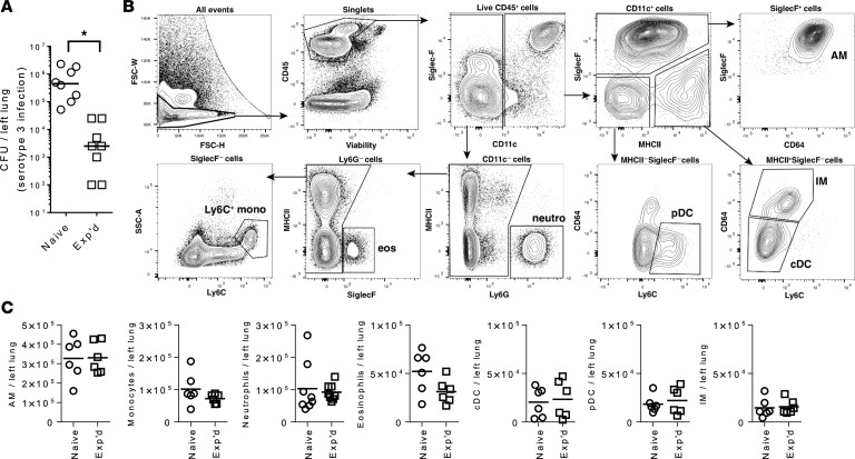
Figure 2. Immunophenotyping of lung myeloid cells 1 month after recovery from mild pneumonia.
(A) Defense against a serotype 3 pneumococcal pneumonia in naive mice and experienced (Exp’d) mice with a history of serotype 19F pneumococcal infections as described in Figure 1. Naive or experienced mice were infected intranasally (i.n.) with 0.5 × 10⁶ to 1 × 10⁶ CFU, and lung bacterial burdens were determined after 24 hours of infection. Asterisk (*) indicates statistically significant (P < 0.05) difference between groups using Mann-Whitney U test. For B and C, mice received intratracheally (i.t.) left lobe serotype 19F infections (Exp’d) or saline instillations (Naive) on days 0 and 7, before left lung lobes were collected at day 37 with no further infections. (B) Gating strategy for myeloid cells in lung single-cell suspensions. Cells were isolated from enzymatically digested mouse lungs, and after the exclusion of doublets, debris, and dead cells, immune cells were identified by CD45 staining. Sequential gating strategy was used to identify myeloid cell subsets. Representative sample from the naive group is shown. (C) Absolute cell counts of identified myeloid cell subsets were obtained and compared. Numbers per mouse were plotted for each cell type, with data collected over 2 independent experiments and each data point representing an individual animal. Groups were compared using 2-tailed Student’s t test, and naive and experienced mice did not significantly differ for any cell type. AM, alveolar macrophages; cDC, conventional dendritic cells; eos, eosinophils; IM, interstitial macrophages; monos, monocytes; neutro, neutrophils; pDC, plasmacytoid dendritic cells.

