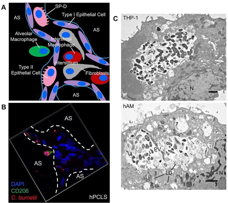
Fig. 2. C. burnetii targets alveolar macrophages in the human lung.
(A) Schematic representation of the alveolar environment and components present. SP-D = surfactant protein D. AS = alveolar space. (B) hPCLS were infected with mCherry-expressing C. burnetii (red) for 72 h, then processed for confocal immunofluorescence microscopy using CD206 antibody (green) to probe for alveolar macrophages and DAPI to stain nuclei (blue). Alveolar macrophages harbor expanded PV containing replicating C. burnetii while other cell types contain very few bacteria and no enlarged PV. (C) THP-1 cells (top) or hAMs (bottom) were infected with C. burnetii for 72 h, then processed for electron microscopy. N = nucleus. LD = lipid droplet. PV = parasitophorous vacuole. Bar, 20 μm. Both macrophage cell types support expanded PV development and robust bacterial growth.

