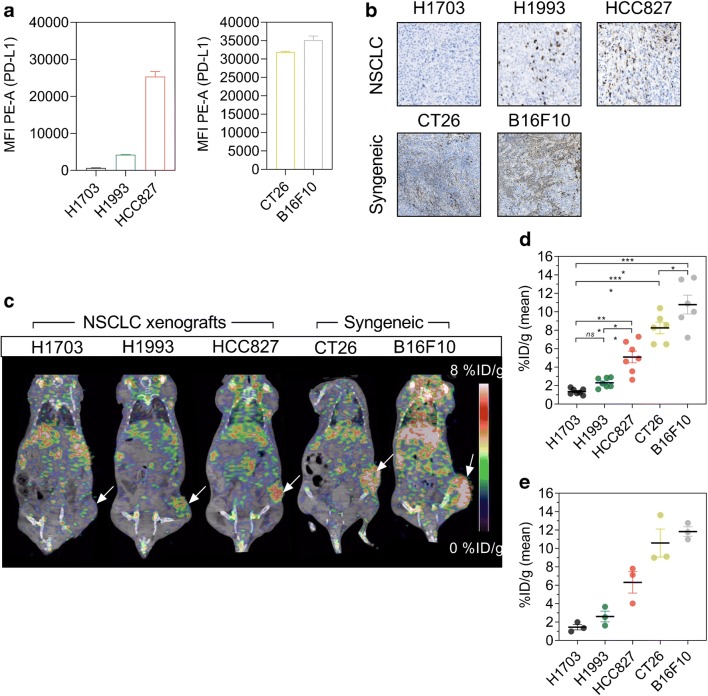
Fig. 3.
Specificity of 89Zr-DFO-6E11 in xenograft and syngeneic mouse tumour models. a Flow cytometric analysis of various cell lines for surface expression of PD-L1 using PE-conjugated anti-human or anti-mouse PD-L1 antibody. Data are presented as a mean of three independent experiments. b Ex vivo immunohistochemical staining of PD-L1 in NSCLC and syngeneic tumour models. c Representative PET/CT images of the H1703, H1993, HCC827 NSCLC xenografts, and CT26 and B16F10 syngeneic tumour models 72 h post-injection of 89Zr-DFO-6E11 illustrating targeting to lymphoid tissue and the tumour (indicated by arrows). d Mean tumour uptake of 89Zr-DFO-6E11 72 h post-injection quantified by PET ROI analysis and expressed as %ID/g (N = 7 for xenograft models, N = 6 for syngeneic models). e Ex vivo uptake in tumours of 89Zr-DFO-6E11 72 h post-injection of the different models measured by gamma counting (N = 3/model). Data are presented as mean ± SEM and the significance levels are indicated by asterisks (*). *p < 0.05, **p < 0.01, ***p < 0.001, ****p < 0.0001. %ID/g, percent injected dose per gram tissue; MFI, median fluorescent intensity; PE-A, phycoerythrin-area; NSCLC, non-small cell lung cancer

