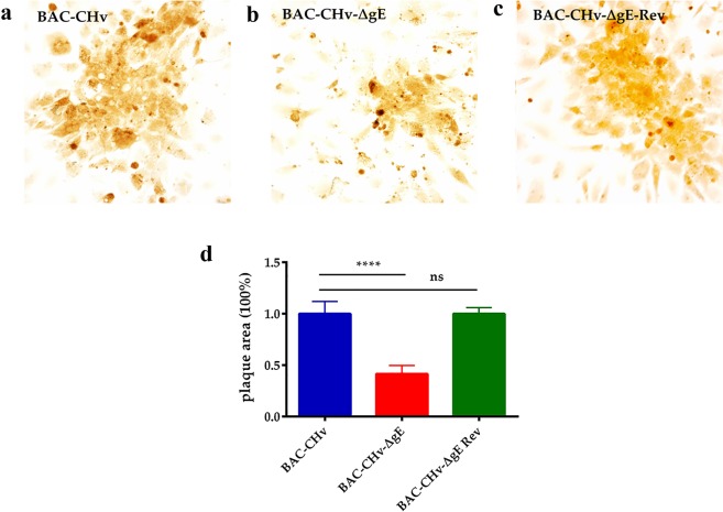
Figure 3.
Plaque morphologies of BAC-CHv, BAC-CHv-∆gE and BAC-CHv-∆gE Rev. DEFs were infected with each virus, and viral plaques were visualized by immunohistochemistry using the polyclonal rabbit anti-CHv antibody at 24 h post-infection. (a) Plaque morphologies of BAC-CHv-infected cells. (b) Plaque morphologies of BAC-CHv-∆gE-infected cells. (c) Plaque morphologies of BAC-CHv-∆gE Rev-infected cells. (d) Statistical analyzation of plaque morphologies of BAC-CHv, BAC-CHv-∆gE and BAC-CHv-∆gE Rev. Error bars represent the standard errors of the means.

