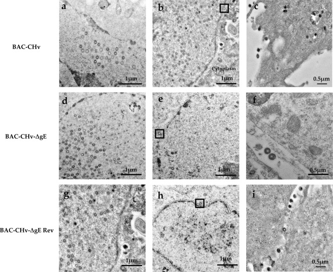
Figure 5.
Ultrastructural morphologies of BAC-CHv, BAC-CHv-∆gE and BAC-CHv-∆gE Rev. DEF cells infected with BAC-CHv, BAC-CHv-∆gE and BAC-CHv-∆gE Rev for electron microscopy at 20 h post-infection were shown. (a) Nucleocapsids accumulated at nuclear in BAC-CHv-infected cells. (b) Primary envelopment process in BAC-CHv infected cells and the process of nucleocapsids de-envelopment at outer nuclear membrane was marked by a black box. (c) BAC-CHv viruses released to extracellular space. (d) Nucleocapsids accumulated at nuclear in BAC-CHv-∆gE infected cells. (e) Primary envelopment process in BAC-CHv-∆gE-infected cells and the process of nucleocapsids de-envelopment at outer nuclear membrane was marked by a black box. (f) BAC-CHv-∆gE viruses released to extracellular space. (g) Nucleocapsids accumulated at nuclear in BAC-CHv-∆gE Rev infected cells. (h) Primary envelopment process in BAC-CHv-∆gE Rev infected cells and the process of nucleocapsids de-envelopment at outer nuclear membrane was marked by a black box. (i) BAC-CHv-∆gE Rev viruses released to extracellular space.

