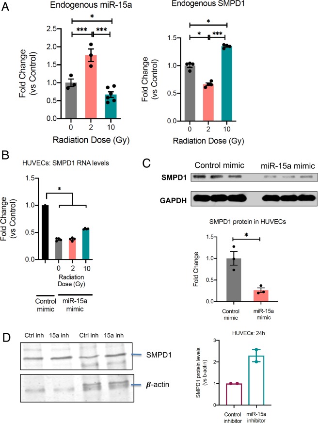
Figure 3.
miR 15a decreases SMPD1 expression in endothelial cells. (A) Reciprocal regulation of endogenous miR-15a (left) and SMPD1 (right) by high vs low dose radiation. HUVECs were irradiated as indicated and RNA was extracted at 18 h post RT. Bars show mean ± SEM of replicates. (B) HUVECs were transfected with either a control mimic or a miR-15a mimic. 24 h later HUVECs were irradiated with the indicated doses. 24 h after radiation, RNA was isolated and qRT-PCR was performed to measure the levels of SMPD1. (C) Cells were lysed at 48 h post transfection and SMPD1 protein levels were measured by western blotting. Lanes show biological replicates and bar graph shows mean band intensity ± SEM of replicates. (D) SMPD1 protein levels in HUVECs 24 h after transfection. Bars show normalized band intensity ± SEM. *P < 0.05, ***P < 0.001 per ANOVA with post hoc Tukey’s test for comparisons between more than 2 groups or two-tailed Student’s T-test for two groups.

