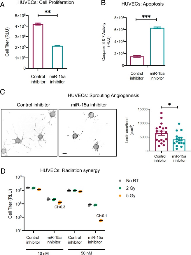
Figure 4.
Inhibition of miR-15a decreases endothelial cell proliferation and enhances cell death. (A) HUVECs were transfected with either a control negative inhibitor or a miR-15a inhibitor. 48 h later proliferation (A) or cell death (B) was measured using a luciferase-based Cell Titer glo assay (A) or Caspase 3 & 7 CasGlo assay (B). (C) Fibrin bead 3D angiogenesis assay. HUVECs were transfected as described in A and were coated onto cytodex beads and allowed to sprout in a fibrin gel in the presence of smooth muscle cells over 5 days. The images show representative beads with angiogenic sprouts stained with Ulex europaeus lectin for each condition. Bars depict mean +SEM of lectin area analyzed across at least 25 beads per group. Scale bar = 100 μm. D) HUVECs were transfected as described in A with the indicated concentrations of either control inhibitor or miR-15a inhibitor. 24 h later, cells were irradiated at the indicated doses. 48 h post irradiation, proliferation was measured using a Cell Titer glo assay. Synergy was calculated using the Chou-Talalay method with combination index < 1 considered synergistic. Bars indicate means ± SEM of 3 technical replicate wells. One of two independent experiments is shown.

