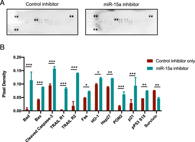
Figure 5.
miR-15a inhibition in endothelial cells drives cell death pathways. (A) Western blot on a membrane array for human apoptosis pathways (Proteome profiler, R&D Systems). HUVECs were transfected with either control inhibitor or miR-15a inhibitor as described in Fig. 4. 48 h later, cells were lysed and the lysates were incubated with a membrane containing duplicate spots with bound antibodies for different apoptotic proteins and developed with SA-HRP per manufacturer’s instructions. (B) Densitometry of duplicate spots from the membrane. Bars show mean + SD. Bars are shown only for proteins that were significantly different between the groups via two-tailed Student’s T-test followed by a Holms-Sidak post-hoc correction for multiple comparisons (Adjusted P-values <0.05). *** denotes adjusted P < 0.0001, ** denotes adjusted P < 0.005, * denotes adjusted P < 0.05.

