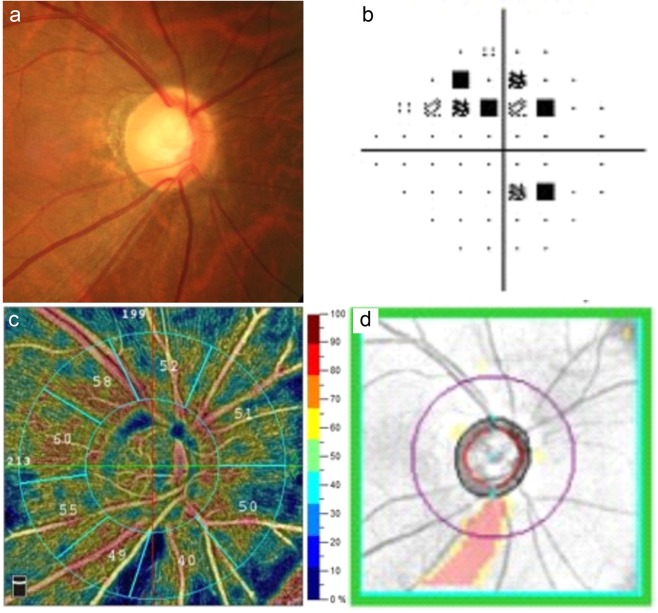
Figure 1.
Images from the right eye of an individual with primary open-angle glaucoma. (a) The optic disc photography shows characteristic vertical elongation of the optic cup accompanied by a loss of inferior temporal circumpapillary retinal nerve fibre layer (cpRNFL). (b) The visual field test shows a superior arcuate defect with a mean deviation of −2.46 decibels. (c) The optical coherence tomography angiography (RTVue XR with AngioVue; version 2018.0.0.14; URL: http://www.optovue.com) shows reduced vessel densities in the inferior temporal sector of radial peripapillary capillary image. (d) The Cirrus optical coherence tomography shows inferior temporal cpRNFL thinning corresponding to that presenting in the optic disc photography.

