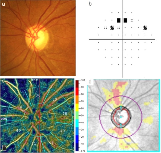
Figure 2.
Images from the left eye of an individual with primary angle-closure glaucoma. (a) The optic disc photography shows characteristic glaucomatous changes. (b) The visual field test shows a superior arcuate defect with a mean deviation of −3.33 decibels. (c) The optical coherence tomography angiography (RTVue XR with AngioVue; version 2018.0.0.14; URL: http://www.optovue.com) shows a generalised reduction of peripapillary vessel densities. (d) The Cirrus optical coherence tomography shows retinal nerve fibre layer thinning corresponding to visual field defects.

