Figure 3.
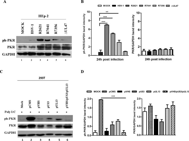
Detection of total PKR and ph-PKR in infection or transfection experiments. (a) HEp-2 cells were mock infected or infected with HSV-1 wild-type virus, R2621 (ΔVHS), R7041 (ΔUs3), R7356 (ΔUL13) mutant viruses at MOI 10 and harvested 24 h p.i. The ∆UL47 virus was used as a viral control. Western blot analysis was performed to detect ph-PKR (Thr-446), PKR and GAPDH expression. (b–d) Band density was determined with the T.I.N.A. program, expressed as fold change over the appropriate housekeeping genes and graphically represented by GraphPad Prism 6. Statistical significance was tested by one-way analysis of variance (ANOVA) (**p < 0.01, and ***p < 0.001). (c) 293T cells were transfected with 1 μg of plasmid DNA according to manufacturer’s instructions. 48 h post transfection the cells were treated with poly I:C (0,01 μg/μl) for an additional 24 h. Western blot analysis was carried out to analyse ph-PKR, PKR and GAPDH expression.

