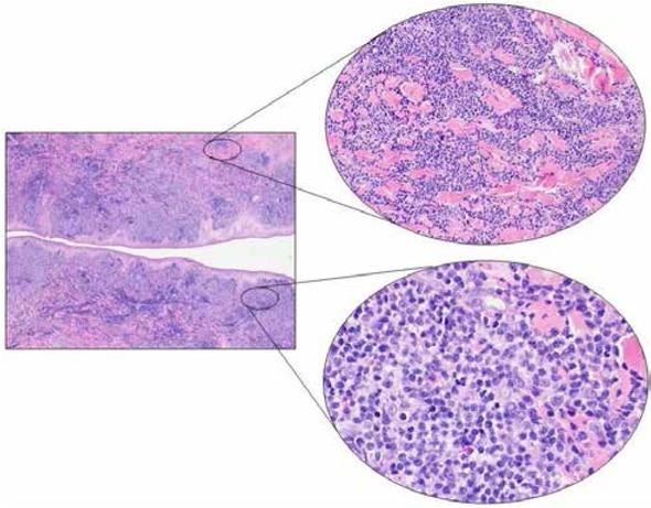
Figure 2.

Microscopic examination showed a dense lymphoid infiltrate involving the reticular dermis, sparing the epidermis and the most superficial papillary dermis (Grenz zone) (Left, H&E, 2x). The lymphoid population showed a diffuse growth pattern peripherally dissociating the collagen bundles. In the deep dermis, at the bottom of the proliferation, a granulomatous reaction was seen with multinucleated giant cells (Upper right, H&E, 20x). The cell population consisted of medium- and large-sized cells with poorly defined cellular borders, slightly eosinophilic cytoplasm, roundish nuclei with irregular chromatin distribution and one or more prominent nucleoli. Several small mature T cells were present in the background (Lower right, H&E, 40x).
Abbreviation: H&E Hematoxylin and Eosin stain
