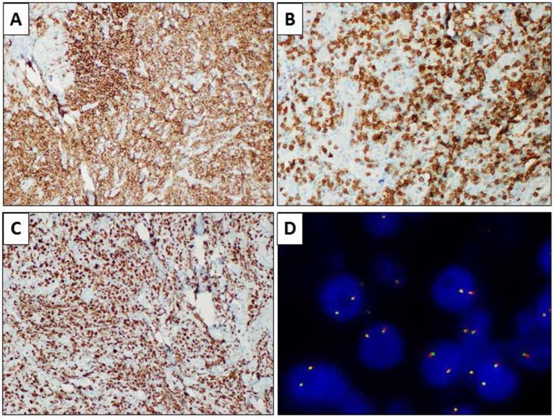
Figure 3.

Immunohistochemical evaluations showing positivity for CD20 (A, immunohistochemical staining, 10x) and negativity for CD3, which was negative in the large neoplastic cells but resulted positive in the T-cell population in the background (B, immunohistochemical staining, 20x). Results for Ki67 were positive in about 90% of the cells (C, immunohistochemical staining, 10x). Fluorescence in-situ hybridization showing wild-type MYC gene (D, break-apart probe FISH).
