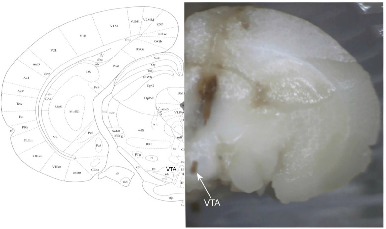
Figure 12.
A photomicrograph of the coronal section indicates the location of the trajectory to and micropipette tip location in the right VTA
In the final session, the implanted rat on the stereotaxic frame received −20 μA of electrical current for 10 min through the recording electrode to deposit microiontophoretic of Pontamine Sky Blue in the recording site. The whole brain was precisely removed and maintained in the 10% phosphate-buffered formalin solution for at least 1 week. The brains were cut by using a microtome to evaluate the location of electrode tips. In the Figure, the location of the VTA is indicated on the histologic image (right) and stereotaxic coordination (left).

