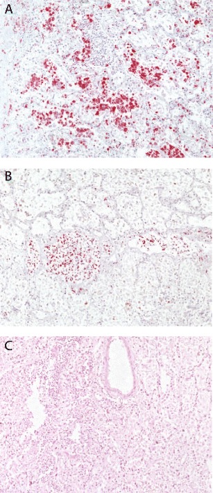
Fig. 1.

Post-mortem histopathological analysis of lung biopsy material obtained from a fatal pregnant case. Lung biopsy material was obtained from a 24-year-old pregnant woman without any known underlying diseases who succumbed to the 2009 H1N1 influenza virus infection after spontaneous expulsion of the fetus [26]. A severe alveolitis with numerous immunohistochemically (red) stained CD68+ macrophages (a) and CD3+ T lymphocytes (b) is observed. At this late stage of infection, influenza virus RNA was not detectable anymore by radioactive in situ hybridization (HE staining) (c). The histochemical stainings were performed according to protocols described before [35]
