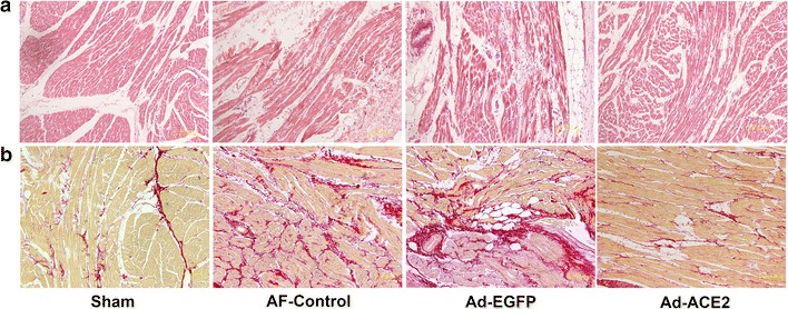
Fig. 1.

Representative images of atrial tissues stained with hematoxylin and eosin (a) and picrosirius red staining (b). Atrial myocyte from sham dogs showed normal composition of sarcomeres distributed throughout the cell, while those from AF-control and Ad-EGFP dogs showed a loss of some contractile materials and abnormal sarcomeres. In addition, extensive interstitial fibrosis, evidenced by picrosirius red stain was found in these tissues. In contrast, these pathologic abnormalities of atrial tissues were attenuated in the Ad-ACE2 group (n = 7)
