Fig. 10.

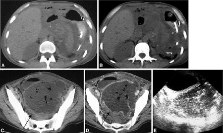
Pelvic abscess. (A–D) CT and (E) US after multivisceral transplantation show a 10-cm pelvic abscess, with thick enhancing walls, containing air bubbles (arrows in C, D). Free abdominal fluid is also visible.

Pelvic abscess. (A–D) CT and (E) US after multivisceral transplantation show a 10-cm pelvic abscess, with thick enhancing walls, containing air bubbles (arrows in C, D). Free abdominal fluid is also visible.