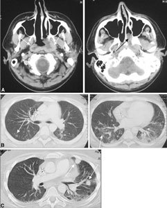
Fig. 12.

PTLD. Large-cell rhinopharyngeal and thoracic malignant lymphoma developed about 1 year after isolated bowel transplantation. A Solid nonhomogeneous tissue of the rhinopharynx wall spreads toward the parapharyngeal and prevertebral spaces (white arrows) and toward the nasal fossa (black arrow). B In the lung, several confluent areas of parenchymal consolidation, some of which are round, are visible (white arrows). C These lesions are larger at 2-month follow-up on CT.
