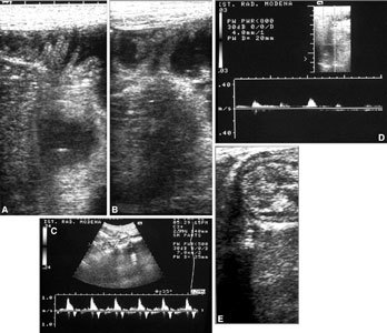
Fig. 4.

US of the bowel in a transplanted patient. A, B Fluid in some intestinal loops permits clearer US identification of intestinal loops. Clearly visible are the wall thickness and valves. Doppler US displays arterial flows (C) in the mesenteric artery and (D) at the level of the intestinal wall. E Normal echoic features and mural stratification of transplanted loops are visible.
