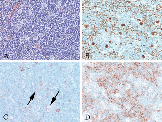
Fig. 2.

NLPHL with rare CD30+ LP cells (case 19). a High power shows large, pale LP cells in a background of small lymphocytes. b LP cells and scattered small B cells show strong nuclear staining for OCT2. c. With antibody to CD30, there are a few faintly positive LP cells (arrows). Also seen are few scattered immunoblasts expressing CD30 more strongly than the LP cells. d The LP cells are present within an irregularly expanded CD21+ follicular dendritic meshwork (b–d immunoperoxidase technique on paraffin sections)
