Abstract
Purpose
Human infections with avian influenza A (H7N9) virus manifested in China in March 2013. The first case infected with H7N9 virus in Beijing involved a family member of a chicken dealer and was reported in April 2013. The clinical and epidemiological characteristics of this case and her parents were examined to illustrate some key traits regarding this novel H7N9 virus.
Methods
The index case was subjected to intensive clinical examination in order to observed the clinical process. Real-time PCR was used to confirm cases infected with H7N9 virus. The index case was administered oseltamivir (45 mg, twice daily) at the early stage of the infection. Sera were collected from the index case and her parents from the onset of illness onwards. The subjects were followed for 4 weeks.
Results
The sera were confirmed by neutralizing antibody tests. The index case’s clinical manifestation progressed quickly. The pharyngeal swab tested positive for influenza A based on the detection of influenza A antigen (rapid influenza diagnostic test) 15 h after the onset of fever and was positive for H7N9 virus. The patient’s temperature dropped to 36.2 °C 18 h after treatment by oseltamivir (32 h after fever). Cough and other symptoms alleviated rapidly. A number of specimens from the environment of this cluster and from the feces specimens tested positive for viral RNA of the H7N9 virus on the fourth day following onset of the index case’s illness. Pharyngeal swabs of the mother tested positive for H7N9 virus twice, but she showed no clinical symptoms. Four weeks after disease onset, the family did not present any clinical symptoms, and the results of the physical examination and blood tests were normal. The mother and the case’s sera had a fourfold increased neutralizing antibody titer.
Conclusion
Early diagnosis and early initiation of the treatment of confirmed infections is the most effective strategy for managing H7N9 virus infection. Human beings exposed to H7N9 virus may develop asymptomatic infection.
Keywords: Avian Influenza A (H7N9), Oseltamivir, Family, Neutralizing antibody titer, Asymptomatic infection
Introduction
In March 2013, patients infected with a novel avian-origin influenza A (H7N9) virus were identified in the Yangtze River Delta of South Eastern China. Influenza A H7N9 is a triple reassortant virus [1]. In contrast to the common clinical features of patients infected with other H7 subtype viruses, those infected with H7N9 rapidly develop lower respiratory tract illness [2–5]. In many cases of H7N9 influenza, the health status of the patients can rapidly deteriorate into severe acute respiratory distress syndrome (ARDS) or multiple organ failure, ultimately progressing to death. The spectrum of H7N9 infections remains unclear as epidemiological data are still limited. Prior 30 May 2013, 132 cases had been diagnosed worldwide, with a case-fatality rate (CFR) of up to 28.03 % (37/132) [6]. An efficient treatment strategy to reduce the CFR is therefore urgently sought in China.
Following notification of the first three confirmed cases of H7N9 infection in the Yangtze River Delta, a hospital-based H7N9 surveillance program was initiated throughout China. All patients presenting with pneumonia caused by an unknown pathogen [definitions: fever (axillary temperature ≥38 °C); radiological characteristics of pneumonia; normal white cell count or leucopenia or lymphopenia; infection of unknown origin] were screened for H7N9 virus [7]. In addition, close contacts were traced and submitted to medical observation.
On 12 April 2013, a 7-year-old girl was confirmed to have been infected with H7N9 virus in Beijing. Her family members were exposed to the same environment. The index case had pneumonia and the mother had an asymptomatic infection. In this report we describe the clinical and epidemiological characteristics of the index case and her family to illustrate a number of key traits regarding this novel H7N9 virus.
Patient
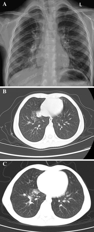
The index case was a 7-year-old girl who was from a family of chicken dealers. She presented with fever, cough and discomfort on 11 April 2013 at 9:00 a.m. She was admitted to Beijing Ditan Hospital, Captial Medical University with a temperature of 38.6 °C. After admission, she underwent radiological and blood tests. Chest radiography showed patchy opacities and edge effusion on bilateral lungs (Fig. 1a).
Fig. 1.

a Chest radiographs of the index case showing patchy opacities and edge effusion on bilateral lungs 5 h after fever. b, c chest computed tomography (CT) scan of the index case: b consolidation in the right lower lobe 15 h after onset of fever, c improvement 3 days after treatment with oseltamivir. The lung shadow of consolidation had became absorbed
After admission, this patient developed fever (maximum temperature 40.2 °C) which could not be brought under control with the administration of acetaminophen (2 ml orally). By 15 h after the fever began, her respiratory rate had risen to 35 breaths per minute and finger oxygen saturation had dropped to 95–96 %. The breath sounds of her right lung became weak, and fine moist rales could be heard, with the intensity of the right lung’s breath sound decreasing. Chest CT scan showed right lower lobe consolidation (Fig. 1b). The pharyngeal swab was positive for influenza A using a rapid influenza diagnostic test based on the presence of influenza A antigens. Subsequently, oseltamivir (45 mg twice daily) was prescribed to the patient. The specimens sent to the Beijing Center for Disease Control and Prevention (CDC) and the Chinese National Influenza Center (CNIC) tested positive for H7N9 virus. The patient’s temperature dropped to 36.2 °C 18 h after the initiation of treatment with oseltamivir. Cough and other symptoms alleviated rapidly. The chest CT scan showed improvement 3 days after disease onset (Fig. 1c).
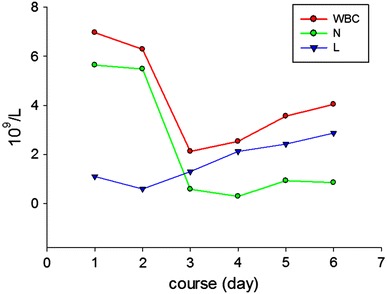
Five hours after the fever began, blood tests showed that the peripheral white blood cell count (WBC) was normal (6.96 × 10⁹/l), although neutrophil (5.64 × 10⁹/l) and lymphocyte counts were depressed (1.1 × 10⁹/l). On the second day after disease onset lymphocyte (0.5 × 10⁹/l), CD4+ T cell (25 cell/μl) and CD8+ T cell (83 cell/μl) counts were significantly depressed. On the third day the lymphocyte count (1.3 × 10⁹/l) began to increase (Fig. 2), and on the fourth day WBC (3.5 × 10⁹/l) and neutrophil (1.1 × 10⁹/l) counts sunk to their lowest respective level. The patient’s platelet count and red blood cell count remained normal during the hospitalization period. The parameters of cardiac enzymes, liver function and arterial blood gas analysis remained normal. The WBC, neutrophil and lymphocyte counts returned to normal 6 days after onset of symptoms.
Fig. 2.

Changes in blood parameters during the course of the illness base on routine blood tests. WBC White blood cell, N neutrophil, L lymphocyte
The index case’s parents, who earned their livelihood by purchasing and selling live chickens, had no clinical symptoms, and examinations of their blood were normal.
Virus isolation and identification
Samples of the patient’s pharyngeal swab, which tested positive for influenza A (see above), were sent to the Beijing CDC and the CNIC for further testing.
RNA was extracted from all specimens using the QIAamp Viral RNA Mini Kit (Qiagen, Venlo, The Netherlands) following the manufacturer’s instructions. Real-time reverse-transcriptase-PCR (RT-PCR) assays were performed to detect the H7N9 virus, relying on specific primers and probes, as described by CNIC. Viral isolation, genome sequencing and phylogenetic analysis of H7N9 viruses were carried out at CNIC [1].
Fifteen hours after the fever began, the pharyngeal swab collected from the index case tested positive for the H7N9 virus by RT-time PCR. About 18 h after the initiation of treatment with oseltamivir, the pharyngeal swab tested positive for influenza A and negative for both H7 and N9. Thirty hours after the initiation of treatment with oseltamivir, all of the pharyngeal swab specimens tested negative for influenza A, H7 and N9. The fecal specimen was available only on the fourth day of the onset of symptoms, and it tested positive for H7N9 virus.
Pharyngeal swabs collected from the index case’s mother tested positive for the H7N9 virus on 12 April and 14 April and were negative after 15 April. At 4 weeks after admission of the index case to the hospital, the father’s pharyngeal swabs tested negative.
Phylogenetic analysis of genome sequences of H7N9 viruses
The influenza virus A/Beijing/1/2013 (H7N9) was isolated from the index case, but the virus could not be isolated from her mother’s specimens or from specimens taken from the patient’s environments.
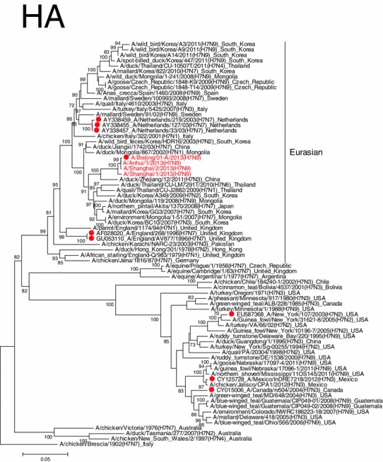
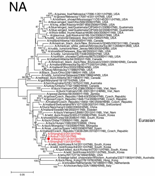
Phylogenetic analysis showed that A/Beijing/1/2013 (H7N9) was highly homologous with A/Shanghai/1/2013 (H7N9) and A/Anhui/1/2013 (H7N9), A/Shanghai/2/2013 (H7N9) and A/Anhui/1/2013 (H7N9) in all eight gene segments (Fig. 3a, b).
Fig. 3.


Phylogenetic tree of eight gene segments the influenza H7N9 virus. Phylogenetic analysis showed that A/Beijing/1/2013 (H7N9) was highly homologous with A/Shanghai/1/2013 (H7N9), A/Shanghai/2/2013 (H7N9) and A/Anhui/1/2013 (H7N9) in all eight gene segments
Seroepidemiologic investigation
We conducted a seroepidemiologic investigation from 12 April to 10 May. Written informed consent was obtained from the patient’s parents. The study was approved by the Ditan Ethics Committee.
Surveillance of the first case began at admission to hospital and continued for 4 weeks. We collected serum on the day of admission to hospital (first day of illness onset), then on post-admission days 7, 14, 21 and 28. The parents of the index case were also tested at the given time-points. The presence of H7N9 neutralizing antibodies within the sera was tested using the microneutralization assay and confirmatory immunoblot assay. Knowledge of the strain of virus and cells used for the neutralization assay would be useful information, as would details of the immunoblot (i.e. antibodies and antigen used). Serological evidence of H7N9 virus infection was defined as an H7N9 neutralizing antibody titer of ≥80 with confirmatory findings from the immunoblot assay. The index case and mother’s sera specimens all possessed a fourfold increased neutralizing antibody titer.
Follow-up of the case family
The parents of the index case were identified as close contacts, and her mother was subsequently diagnosed with asymptomatic infection. All of her father’s specimens that were collected during the period of medical observation tested negative for H7, N9 and influenza A, and he did not develop any influenza-like symptoms during this period.
The index case was discharged on post-admission day 7. Two weeks after the illness onset (1 week after discharge), the index case and her parents were followed-up. They did not present any clinical symptoms, and the results of physical examination and blood tests were normal.
Discussion
This report provides a new insight into the characteristics of H7N9 infection, including the strategy of treatment, spectrum of this disease, viral shedding, and the source of transmission.
The gene sequences of the virus isolated from the index case had a high homology with those from the cases of H7N9 infection detected in South East China (Fig. 3), suggesting that this virus would have a similar virulence as those viruses leading to severe symptoms and death in patients reported previously [1]. The time-frame of patient treatment these cases was upon admission (index case in Beijing) and 5 h and 6–8 days after onset of symptoms (index cases in South East China); antiviral agents were administered to the cases at 15 h and 7–8 days after symptom onset, respectively.
Within 5 h onset of symptoms, the index case developed pneumonia, persistent high fever, local lobe consolidation and remarkable lymphopenia. Given the rapid progression of the illness in the previously reported H7N9 infected patients [1], ARDS was expected to occur if the causative agent and appropriate cause of treatment were not identified rapidly. A rapid influenza A antigen test was performed 15 h after the fever began, and the positive result precipitated early application of oseltamivir. Maximal plasma concentrations of oseltamivir carboxylate (OC), the active metabolite of oselatamivir, occur approximately 3–4 h after administration. The plasma half-life of OC is 6–10 h, which enables a twice-daily dosing regimen [8, 9]. Our index case recovered rapidly and did not develop ARDS, an outcome which may be attributed to the early administration of oseltamivir (15 h after onset of fever). The pharyngeal swab collected from the index case tested negative 30 h after treatment with oseltamivir.
The specimen of feces from the index case tested positive for H7N9 viral RNA 4 days after the onset of symptoms, indicating that this virus was able to cause enteric infection, such as H5N1 virus [10–12]. This finding suggests that the prevention and control of enteric transmission should be pursued in the treatment and management of patients infected with the H7N9 virus.
Among the family of individuals infected with the H7N9 virus in Beijing, one asymptomatic infection was observed, which was the first reported case of asymptomatic infection in China since this virus was identified. We report here the first documented study on asymptomatic infection with avian influenza virus that was identified by the detection of viral RNA rather than on the results of retrospective assays of antibodies against avian influenza virus [13–16].
Although the family members of the index case were all exposed to asymptomatic chickens infected with H7N9 virus, they presented with distinct outcomes. The daughter developed pneumonia, the mother remained asymptomatic, and the father remained free of infection.
In addition, the presence of Q226L in the HA protein and the substitution of E627K in the PB2 protein found in the H7N9 virus reported by Gao et al. indicates that this virus may have an increased ability to infect mammals [1, 17–19]. Taken together, these results suggest that humans may have a higher susceptibility to H7N9 viruses, which poses a serious public health concern. Our experience with the index case suggests that early diagnosis and treatment is the most effective strategy to limit H7N9 virus infection. This is first report to document the different clinical presentations within a family of infected and exposed individuals.
Conflict of interest
None.
Footnotes
R. Song, X. Pang, P. Yang, Y. Shu, Y. Zhang, Q. Wang, Z. Chen, J. Liu, and X. Li contributed equally to this work.
Contributor Information
R. Song, Phone: +86-10-84323039, Email: songruii@hotmail.com
Y. Deng, Email: dengyingbjcdc@126.com
X. Li, Phone: +86-10-84322585, Email: ditanlxw@163.com
References
- 1.Gao RB, Cao B, Hu Y, et al. Human infection with a novel avian-origin influenza A (H7N9) virus. N Engl J Med. 2013;368:1888–1897. doi: 10.1056/NEJMoa1304459. [DOI] [PubMed] [Google Scholar]
- 2.Fouchier RA, Schneeberger PM, Rozendaal FW, et al. Avian influenza A virus (H7N7) associated with human conjunctivitis and a fatal case of acute respiratory distress syndrome. Proc Natl Acad Sci USA. 2004;101:1356–1361. doi: 10.1073/pnas.0308352100. [DOI] [PMC free article] [PubMed] [Google Scholar]
- 3.Koopmans M, Wilbrink B, Conyn M, et al. Transmission of H7N7 avian influenza A virus to human beings during a large outbreak in commercial poultry farms in the Netherlands. Lancet. 2004;363:587–593. doi: 10.1016/S0140-6736(04)15589-X. [DOI] [PubMed] [Google Scholar]
- 4.Kurtz J, Manvell RJ, Banks J. Avian influenza virus isolated from a woman with conjunctivitis. Lancet. 1996;348:901–902. doi: 10.1016/S0140-6736(05)64783-6. [DOI] [PubMed] [Google Scholar]
- 5.Editorial team. Avian influenza A/(H7N2) outbreak in the United Kingdom. Euro Surveill. 2007;12: E070531.2. [PubMed]
- 6.World Health Organization/Global Influenza Program. Cumulative number of confirmed human cases for avian influenza A(H5N1) reported to WHO, 2003–2013. Available at: http://www.who.int/influenza/human_animal_interface/influenza_h7n9/08_ReportWebH7N9Number.pdf. Accessed 30 May 2013.
- 7.Guideline on prevention and control of avian influenza A (H7N9). Available at: http://www.gov.cn/gzdt/2013-04/11/content_2374926.htm. Accessed 3 Apr 2013.
- 8.Smith JR, Rayner CR, Donner B, Wollenhaupt M, Klumpp K, Dutkowski R. Oseltamivir in seasonal, pandemic, and avian influenza: a comprehensive review of 10-years clinical experience. Adv Ther. 2011;28:927–959. doi: 10.1007/s12325-011-0072-7. [DOI] [PMC free article] [PubMed] [Google Scholar]
- 9.Soepandi PZ, Burhan E, Mangunnegoro H, et al. Clinical course of avian influenza A (H5N1) in patients at the Persahabatan Hospital, Jakarta, Indonesia, 2005–2008. Chest. 2010;138:665–673. doi: 10.1378/chest.09-2644. [DOI] [PMC free article] [PubMed] [Google Scholar]
- 10.Buchy P, Mardy S, Vong S, et al. Influenza A/H5N1 virus infection in humans in Cambodia. J Clin Virol. 2007;39:164–168. doi: 10.1016/j.jcv.2007.04.010. [DOI] [PubMed] [Google Scholar]
- 11.Uiprasertkul M, Puthavathana P, Sangsiriwut K, et al. Influenza A H5N1 replication sites in humans. Emerg Infect Dis. 2005;11:1036–1041. doi: 10.3201/eid1107.041313. [DOI] [PMC free article] [PubMed] [Google Scholar]
- 12.de Jong MD, Bach VC, Phan TQ, et al. Fatal avian influenza A (H5N1) in a child presenting with diarrhea followed by coma. N Engl J Med. 2005;352:686–691. doi: 10.1056/NEJMoa044307. [DOI] [PubMed] [Google Scholar]
- 13.Vong S, Ly S, Van Kerkhove MD, et al. Risk factors associated with subclinical human infection with avian influenza A (H5N1) virus—Cambodia, 2006. J Infect Dis. 2009;199:1744–1752. doi: 10.1086/599208. [DOI] [PubMed] [Google Scholar]
- 14.Schultsz C, Nguyen VD, le Hai T, et al. Prevalence of antibodies against avian influenza A (H5N1) virus among cullers and poultry workers in Ho Chi Minh City, 2005. PLoS ONE. 2009;4:e7948. doi: 10.1371/journal.pone.0007948. [DOI] [PMC free article] [PubMed] [Google Scholar]
- 15.Meijer A, Bosman A, van de Kamp EE, et al. Measurement of antibodies to avian influenza virus A(H7N7) in humans by hemagglutination inhibition test. J Virol Methods. 2006;132:113–120. doi: 10.1016/j.jviromet.2005.10.001. [DOI] [PubMed] [Google Scholar]
- 16.Di Trani L, Porru S, Bonfanti L, et al. Serosurvey against H5 and H7 avian influenza viruses in Italian poultry workers. Avian Dis. 2012;56:1068–1071. doi: 10.1637/10184-041012-ResNote.1. [DOI] [PubMed] [Google Scholar]
- 17.Herfst S, Schrauwen EJ, Linster M, et al. Airborne transmission of influenza A/H5N1 virus between ferrets. Science. 2012;336:1534–1541. doi: 10.1126/science.1213362. [DOI] [PMC free article] [PubMed] [Google Scholar]
- 18.Imai M, Watanabe T, Hatta M, et al. Experimental adaptation of an influenza H5 HA confers respiratory droplet transmission to a reassortant H5HA/H1N1 virus in ferrets. Nature. 2012;486:420–428. doi: 10.1038/nature10831. [DOI] [PMC free article] [PubMed] [Google Scholar]
- 19.Labadie K, Dos Santos Afonso E, Rameix-Welti MA, et al. Host-range determinants on the PB2 protein of influenza A viruses control the interaction between the viral polymerase and nucleoprotein in human cells. Virology. 2007;362:271–282. doi: 10.1016/j.virol.2006.12.027. [DOI] [PubMed] [Google Scholar]


