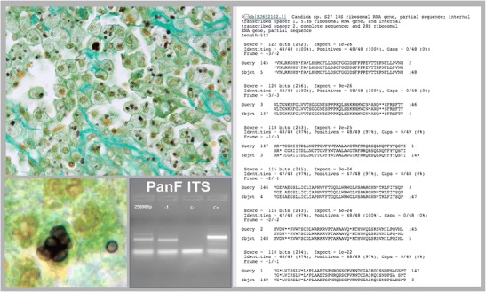
Fig. 1.

Biopsy showing a subcutaneous granuloma. The Grocott stain shows small yeast-like cells compatible with Histoplasma capsulatum and Candida glabrata (on the left) (original magnification × 40, inset × 100). Panfungal PCR that amplifies the internal transcriber spacer (ITS-1) region of the rDNA gene (panel to the left) that after the sequence matched with Candida spp. (panel on the right). The final diagnosis was Candida glabrata
