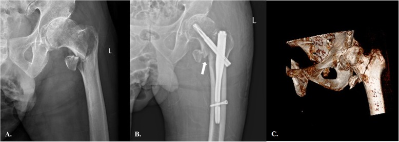
Fig. 2.
a The preoperative anteroposterior (AP) radiograph showing an apparently simple AO-31A1.3 (2018 version) pertrochanteric fracture. b Following fixation with PFNA, there was an obvious inferior gap at the basicervical region (white arrow) and moderate valgus deformity. The lucency around helix indicates a micro-motion and instability of the fracture-implantation complex. c However, the preoperative 3D-CT revealed that this case was a basicervical trochanteric fracture variant. On preoperative radiograph, the external rotation of the femoral shaft made the fracture line assessment and classification incorrectly

