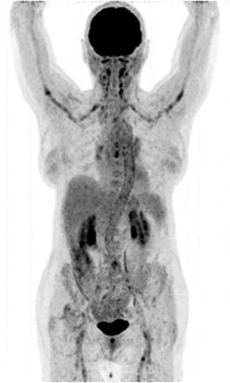
Fig. 8.

Giant cell arteritis. Note the intense, relatively homogeneous [18F]FDG accumulation in the carotid and subclavian arteries, as well as in the aortic arch, thoracic and abdominal aorta, and the iliac and femoral arteries. Imaging was performed about 1 h after injection of tracer
