
Fig. 3.
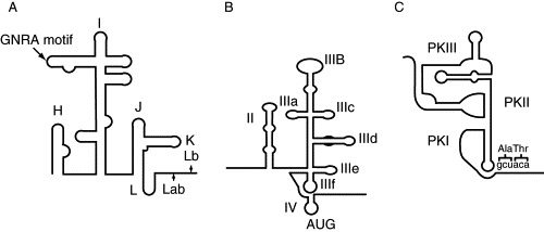
IRES secondary structures. (A) Secondary structure model of the FMDV IRES, a type II picornaviral IRES. The FMDV IRES directs translation initiation from both AUG Lab and AUG Lb sites. Most picornaviral IRESs use a single initiation codon, analogous to FMDV AUG10 for type II and AUG11 for type I IRESs. (B) A structural model of the hepatitis C virus (HCV) IRES. The basal part of domain III is involved in 40S ribosomal subunit binding, and the apical loops of this domain in binding to eIF3. (C). The cricket paralysis virus (CrPV) IRES adopts a triple pseudoknot structure. PKI mimics a tRNA in the ribosomal P site, allowing initiation to occur at a GCU codon in the A site.