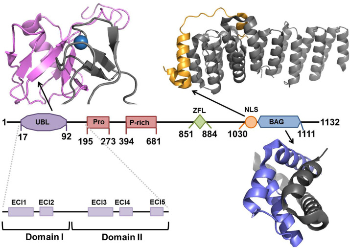
Fig. 3.
Schematic representation of BAG6 showing domain boundaries and solved structures. UBL domain (residues 17–92, purple) in complex with E3 ligase RNF126 (residues 1–40, gray) from PDB: 2N9P (Krysztofinska et al., 2016); NLS (residues 1008–1050, orange) and TRC35 (residues 23–305, gray) from PDB: 6AU8 (Mock, Xu, Ye, & Clemons, 2017); BAG domain (blue) in complex with UBL4A (residues 95–147, gray), from PDB: 4X86 (Kuwabara et al., 2015).

